|
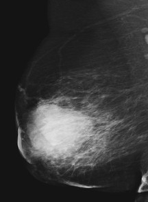
According to BI-RADS, a mass is defined as a space occupying lesion seen in at least two different projections. If a potential mass is seen in only a single projection it should be called 'Asymmetry' or 'Asymmetric Density' until its three-dimensionality is confirmed. Masses have different density (fat containing masses, low density, isodense, high density), different margins (circumscribed, microlobular, obscured, indistinct, spiculated) and different shape (round, oval, lobular, irregular). Round and oval shaped masses with smooth and circumscribed margins usually indicate benign changes. On the other hand, a malignant mass usually has a spiculated, rough and blurry boundary. N.R. Mudigonda, R.M. Rangayyan, J.E.L. Desautels, Detection of Breast Masses in Mammograms by Density Slicing and Texture Flow-Field Analysis, IEEE Transactions on Medical Imaging, Vol. 20, No. 12, 2001, pp. 1215-1227 N. Karssemeijer, J.D.M. Otten, J.D.M., A.L.M. Verbeek, J.H. Groenewoud, H.J. De Koning, J.H.C.L. Hendriks, R. Holland, Computer-Aided Detection versus Independent Double Reading of Masses on Mammograms, Radiology, Vol. 227, No. 1, 2003, pp. 192-200 H.D. Cheng, X.J. Shi, R. Min, L.M. Hu, X.P. Cai, H.N. Du, Approaches for Automated Detection and Classification of Masses in Mammograms, Pattern Recognition, Vol. 39, No. 4, 2006 pp. 646-668 B. Zheng, Y.-H. Chang, D. Gur, Computerized Detection of Masses in Digitized Mammograms Using Single-Image Segmentation and a Multilayer Topographic Feature Analysis, Academic Radiology, Vol. 2, No. 11, 1995, pp. 959-966 S. Paquerault, N. Petrick, H.-P. Chan, B. Sahiner, M.A. Helvie, Improvement of Computerized Mass Detection on Mammograms: Fusion of Two-View Information, Medical Physics, Vol. 29, No. 2, 2002, pp. 238-247 I. Christoyianni, E. Dermatas, G. Kokkinakis, Fast Detection of Masses in Computer-Aided Mammography, IEEE Signal Processing Magazine, Vol. 17, No. 1, 2000, pp. 54-64 S. van Engeland, N. Karssemeijer, Combining Two Mammographic Projections in a Computer Aided Mass Detection Method, Medical Physics, Vol. 34, No. 3, 2007, pp. 898-905 N. Petrick, H.-P. Chan, D. Wei, B. Sahiner, M.A. Helvie, D.D. Adler, Automated Detection of Breast Masses on Mammograms Using Adaptive Contrast Enhancement and Texture Classification, Medical Physics, Vol. 23, No. 10, 1996, pp. 1685-1696 G.D. Tourassi, R. Vargas-Voracek, D.M. Catarious Jr., C.E. Floyd, Computer-Assisted Detection of Mammographic Masses: A Template Matching Scheme Based on Mutual Information, Medical Physics, Vol. 30, No. 8, 2003, pp. 2123-2130 B. Sahiner, H.-P. Chan, N. Petrick, M.A. Helvie, L.M. Hadjiiski, Improvement of Mammographic Mass Characterization Using Spiculation Measures and Morphological Features, Medical Physics, Vol. 28, No 7, 2001, pp. 1455-1465 W.E. Polakowski, D.A. Cournoyer, S.K. Rogers, M.P. Desimio, D.W. Ruck, J.W. Hoffmeister, R.A. Raines, Computer-Aided Breast Cancer Detection and Diagnosis of Masses Using Difference of Gaussians and Derivative-Based Feature Saliency, IEEE Transactions on Medical Imaging, Vol. 16, No. 6, 1997, pp. 811-819 back to top
|  |
|
Calcifications are deposits of calcium in breast tissue. Benign calcifications are usually larger and coarser with round and smooth contours. Malignant calcifications tend to be numerous, clustered, small, varying in size and shape, angular, irregularly shaped and branching in orientation. H.D. Cheng, X. Cai, X. Chen, L. Hu, X. Lou, Computer-Aided Detection and Classification of Microcalcifications in Mammograms: A Survey, Pattern Recognition, Vol. 36, No. 12, 2003, pp. 2967-2991 N. Karssemeijer, Stochastic Model for Automated Detection of Calcifications in Digital Mammograms, Image and Vision Computing, Vol. 10, No. 6, 1992, pp. 369-375 H.-P. Chan, K. Doi, C.J. Vyborny, R.A. Schmidt, C.E. Metz, K.L. Lam, T. Ogura, Y. Wu, H. MacMahon, Improvement in Radiologists' Detection of Clustered Microcalcifications on Mammograms. The Potential of Computer-Aided Diagnosis, Investigative Radiology, Vol. 25, No. 10, 1990, pp. 1102-1110 S. Yu, L. Guan, A CAD System for the Automatic Detection of Clustered Microcalcifications in Digitized Mammogram Films, IEEE Transactions on Medical Imaging, Vol. 19, No. 2, 2000, pp. 115-126 W. Zhang, K. Doi, M.L. Giger, R.M. Nishikawa, R.A. Schmidt, An Improved Shift-Invariant Artificial Neural Network for Computerized Detection of Clustered Microcalcifications in Digital Mammograms, Medical Physics, Vol. 23, No. 4, 1996, pp. 595-601 R.N. Strickland, H. Hahn II, Wavelet Transforms for Detecting Microcalcifications in Mammograms, IEEE Transactions on Medical Imaging, Vol. 15, No. 2, 1996, pp. 218-229 I. El-Naqa, Y. Yang, M.N. Wernick, N.P. Galatsanos, R.M. Nishikawa, A Support Vector Machine Approach for Detection of Microcalcifications, IEEE Transactions on Medical Imaging, Vol. 21, No. 12, 2002, pp. 1552-1563 H.-D. Cheng, Y.M. Lui, R.I. Freimanis, A Novel Approach to Microcalcification Detection Using Fuzzy Logic Technique, IEEE Transactions on Medical Imaging, Vol. 17, No. 3, 1998, pp. 442-450 M. Kallergi, Computer-Aided Diagnosis of Mammographic Microcalcification Clusters, Medical Physics, Vol. 31, No. 2, 2004, pp. 314-326 T.C. Wang, N.B. Karayiannis, Detection of Microcalcifications in Digital Mammograms Using Wavelets, IEEE Transactions on Medical Imaging, Vol. 17, No. 4, 1998, pp. 498-509 back to top
|  |
|
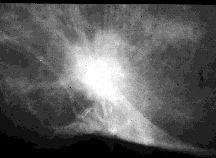
Architectural distortion is defined as distortion of the normal architecture with no definite mass visible, including spiculations radiating from a point and focal retraction or distortion at the edge of the parenchyma. Architectural distortion of breast tissue can indicate malignant changes especially when integrated with visible lesions such as mass, asymmetry or calcifications. Architectural distortion can be classified as benign when including scar and soft-tissue damage due to trauma. J.A. Baker, E.L. Rosen, J.Y. Lo, E.I. Gimenez, R. Walsh, M.S. Soo, Computer-Aided Detection (CAD) in Screening Mammography: Sensitivity of Commercial CAD Systems for Detecting Architectural Distortion, American Journal of Roentgenology, Vol. 181, No. 4, 2003, pp. 1083-1088 R.M. Rangayyan, F.J. Ayres, Gabor Filters and Phase Portraits for the Detection of Architectural Distortion in Mammograms, Medical and Biological Engineering and Computing, Vol. 44, No. 10, 2006, pp. 883-894 G.D. Tourassi, C.E. Floyd Jr., Performance Evaluation of an Information-Theoretic CAD Scheme for the Detection of Mammographic Architectural Distortion, Proceedings of SPIE - The International Society for Optical Engineering 5370 I, 2004, pp. 59-66 G.D. Tourassi, N.H. Eltonsy, A.S. Elmaghraby, C.E. Floyd Jr., Detection of Architectural Distortion in Mammograms Using Fractal Analysis, Progress in Biomedical Optics and Imaging - Proceedings of SPIE 5747 (II), art. no. 97, 2005, pp. 930-938 back to top
|  |
|
Asymmetry of breast parenchyma between the two sides is useful sign for detecting primary breast cancer. Bilateral asymmetries of concern are those that are changing or enlarging or new, those that are palpable and those that are associated with other findings, such as microcalcifications or architectural distortion. If a palpable thickening or mass corresponds to an asymmetric density, the density is regarded with a greater degree of suspicion for malignancy. F.-F. Yin, M.L. Giger, K. Doi, C.J. Vyborny, R.A. Schmidt, Computerized Detection of Masses in Digital Mammograms: Automated Alignment of Breast Images and its Effect on Bilateral-Subtraction Technique, Medical Physics, Vol. 21, No. 3, 1994, pp. 445-452 R.J. Ferrari, R.M. Rangayyan, J.E.L. Desautels, A.F. Frère, Analysis of Asymmetry in Mammograms via Directional Filtering with Gabor Wavelets, IEEE Transactions on Medical Imaging, Vol. 20, No. 9, 2001, pp. 953-964 T.-K. Lau, W.F. Bischof, Automated Detection of Breast Tumors Using the Asymmetry Approach, Computers and Biomedical Research, Vol. 24, No. 3, 1991, pp. 273-295 A.J. Méndez, P.G. Tahoces, M.J. Lado, M. Souto, J.J. Vidal, Computer-Aided Diagnosis: Automatic Detection of Malignant Masses in Digitized Mammograms, Medical Physics, Vol. 25, No. 6, 1998, pp. 957-964 F. Georgsson, Differential Analysis of Bilateral Mammograms, International Journal of Pattern Recognition and Artificial Intelligence, Vol. 17, No. 7, 2003, pp. 1207-1226 back to top | 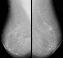 |
Breast abnormalities that can indicate breast cancer are masses, calcifications, architectural distortion and bilateral asymmetry. Here we give a brief description of each of these abnormalities together with the most representative scientific papers published in high impact factor journals. One of the criteria for the selection of papers was their relevance and the number of citations according to SCOPUS database.
